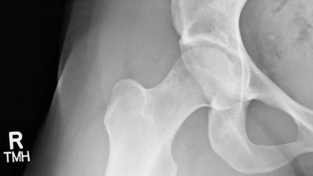
The Dunn view is a specialized X-ray positioning technique used to evaluate the hip joint, particularly in cases of femoroacetabular impingement (FAI) or other structural abnormalities. Key Difference from the Frog-Leg View While the frog-leg lateral view superimposes the greater and lesser trochanters, the Dunn view aims to keep them separate for better visualization of […]
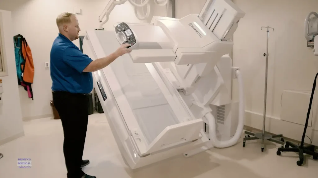
Upgrading to the X-1 radiography system from Brown’s Medical Imaging has brought major improvements to both patient care and imaging efficiency. Better Access for All Patients With an increased deck height range, the table can be lowered significantly, making it much easier to accommodate: Wheelchair patients, who can now transfer more comfortably and safely. Sterile […]
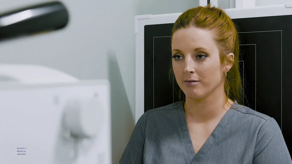
At True Roots Chiropractic, the mission is clear: remove nerve interference so the body can function at its optimal level. One of the best tools for achieving that goal is having the ability to “look inside” and see what’s going on beneath the surface — and that’s where digital X-ray technology comes in. Better Data […]
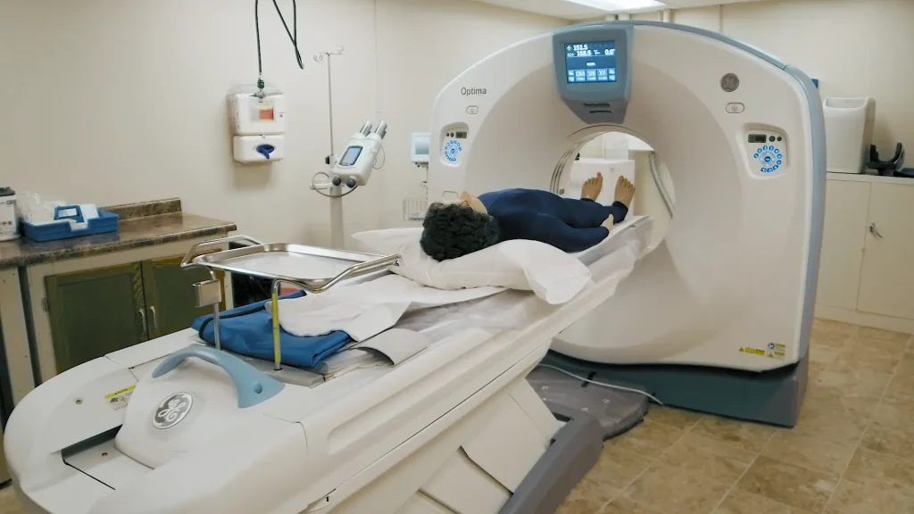
In a medical emergency, every minute matters. In one recent stroke case, the facility was able to scan and diagnose the patient within minutes using their in-house CT system. This was a major improvement compared to previous delays when patients had to be sent to another facility for imaging. Keeping Patients — and Revenue — […]
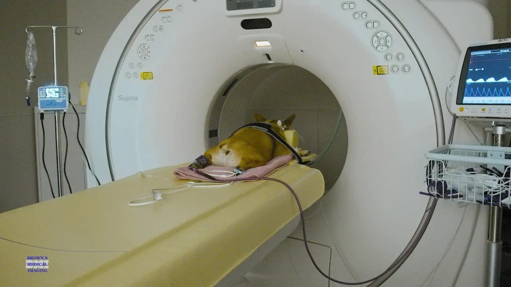
At our emergency and specialty veterinary clinic, we often handle the cases that general practice partners refer when a pet’s needs go beyond routine care. These complex cases require precise diagnostics — and that’s where CT imaging plays a vital role. Seeing Inside Without Being Invasive CT allows our team to view a pet’s internal […]
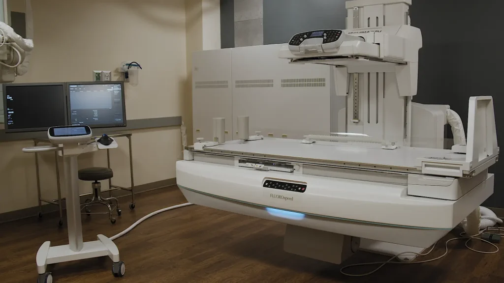
After nearly two decades with the same equipment, this department is stepping into a new era of patient care with a state-of-the-art imaging table and system from Brown’s Medical Imaging. Designed for Patient Comfort and Safety The old table was high off the ground, making it difficult for older patients or those with mobility challenges […]
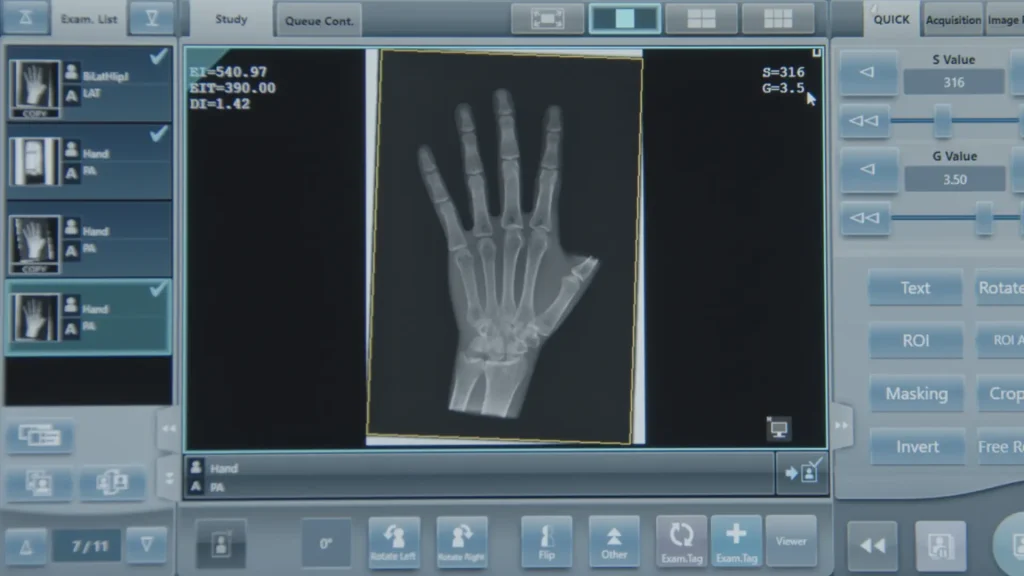
With digital imaging, brightness and contrast are no longer controlled solely by exposure factors. Modern systems post-process images to deliver consistent density and contrast, even when exposure factors vary. While this provides greater latitude, it also means a radiographer can’t rely on image appearance alone to judge whether an exposure was too high or too […]
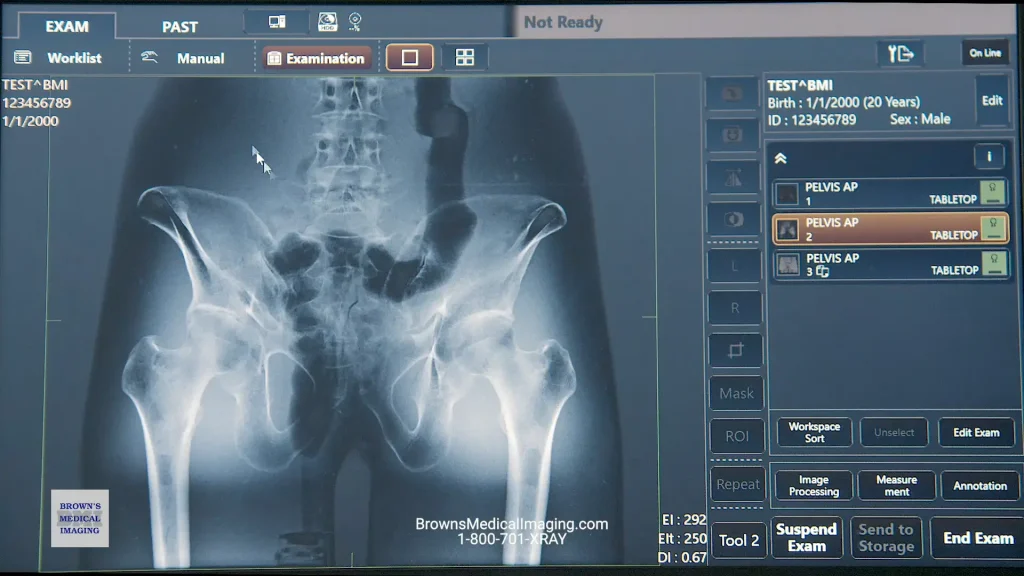
The Canon control software is designed to make digital radiography more efficient, intuitive, and customizable. With easy patient registration, powerful image tools, and built-in workflow shortcuts, technologists can work faster while maintaining high image quality. Efficient Patient Management Exam Tab: Register patients from a worklist or enter them manually if a worklist isn’t available. Pass […]
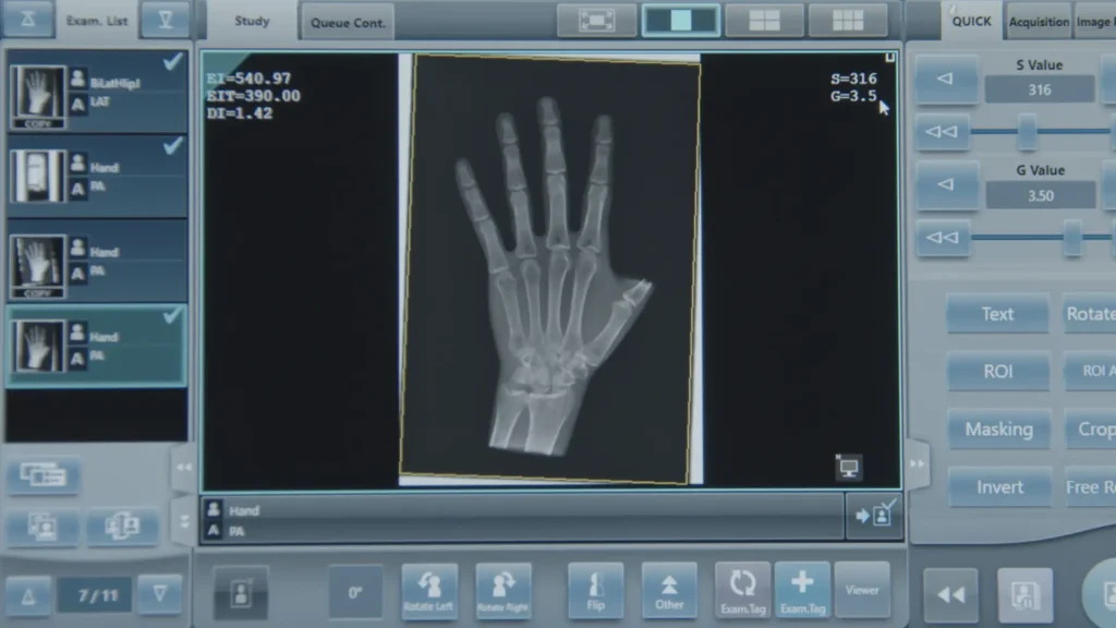
The Konica Minolta RC7 is designed to give technologists powerful tools for efficient workflow, image optimization, and streamlined patient care. While the software is robust, most work is done in the Order List, where exams are selected and performed. From there, the RC7 makes it easy to manage studies, adjust images, and send them to […]
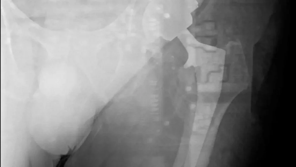
In the effort to make digital radiography (DR) detectors lighter and easier to handle, manufacturers have reduced the amount of internal shielding on the backside of detectors. While this improves portability, it can also increase the risk of backscatter radiation artifacts — especially during challenging positioning angles and high-exposure exams. A radiograph affected by backscatter […]
