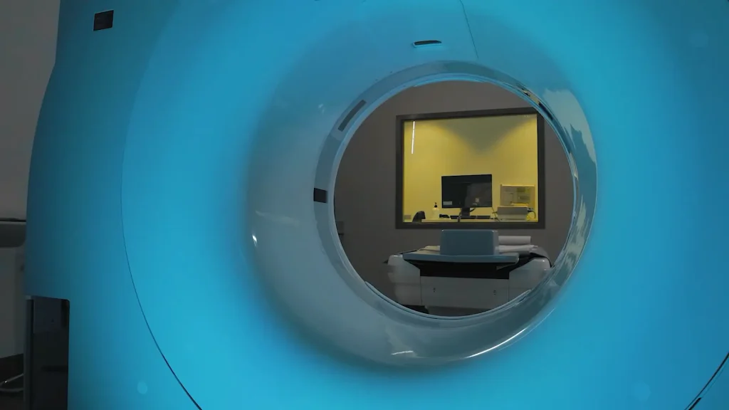
Switching from an older CT scanner to a Fujifilm system has completely transformed the imaging workflow. What once required multiple post-processing steps is now fully automated, saving time for technologists and creating a smoother experience for patients. Automated Efficiency With the Fujifilm system, once the exam is complete, the scanner automatically performs all reconstructions and […]
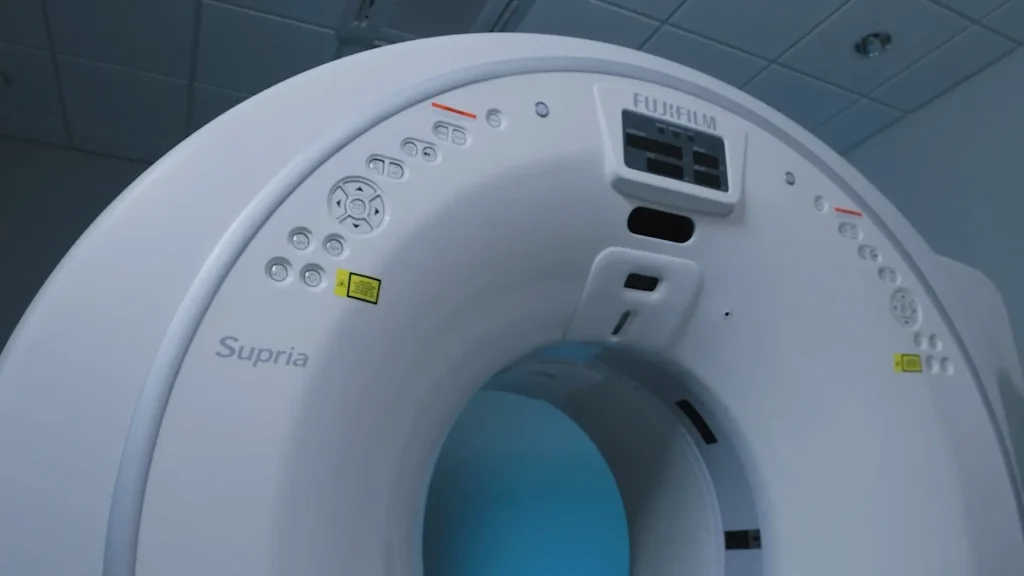
Ear, Nose and Throat of Utah is home to 13 physicians, including specialists in throat cancer, otology and neurology, and pediatric ENT care. With a wide range of patients—from those with sinus issues to allergy concerns—having CT imaging readily available in-house is a major advantage. Why In-House Imaging Matters Instead of sending patients to a […]

At just 15 years old, a CT scan unexpectedly revealed a breast cancer diagnosis. While the news was life-changing, the experience also sparked an interest in medical imaging that would shape a future career. How the Experience Inspired a Career Following the CT scan, the journey continued with ultrasounds and a mammogram. Experiencing firsthand how […]
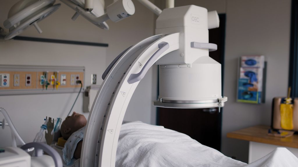
Radiography students often assist with surgeries or invasive procedures using a C-arm during clinical rotations—a setting that can feel high-pressure. Having access to a refurbished C-arm from Brown’s Medical Imaging on campus allows students to learn and practice in a low-stakes environment before they step into real-world situations. How It Benefits Students Safe Learning Space […]
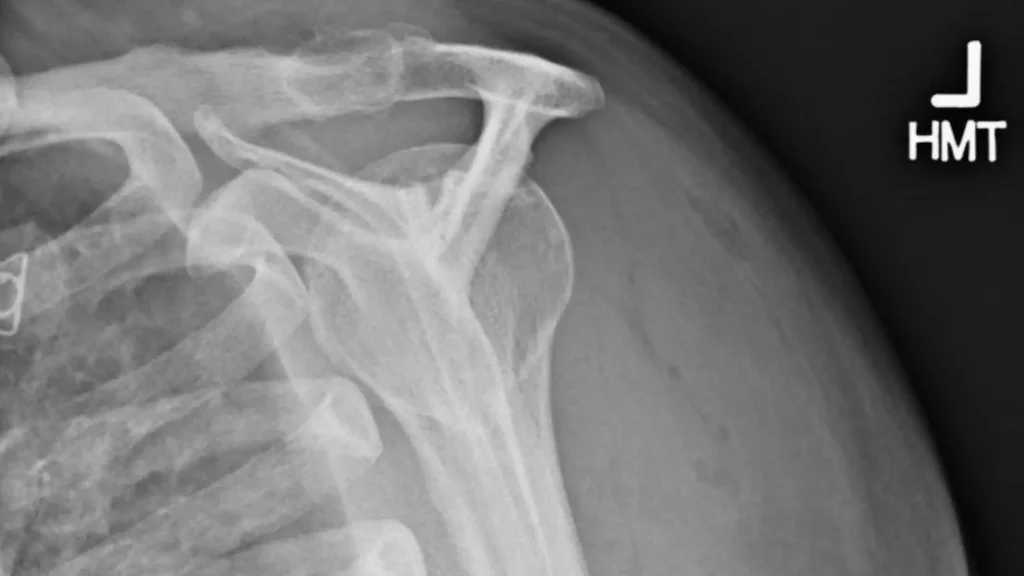
The Y view, or outlet view, is a valuable projection for evaluating the acromion’s shape and overall shoulder alignment. This positioning technique ensures a clear, diagnostic image while maintaining patient comfort and minimizing repeats. Key Positioning Steps Set the Angulation – Use a 20-degree caudal angle to highlight the acromion and surrounding anatomy. Find the […]
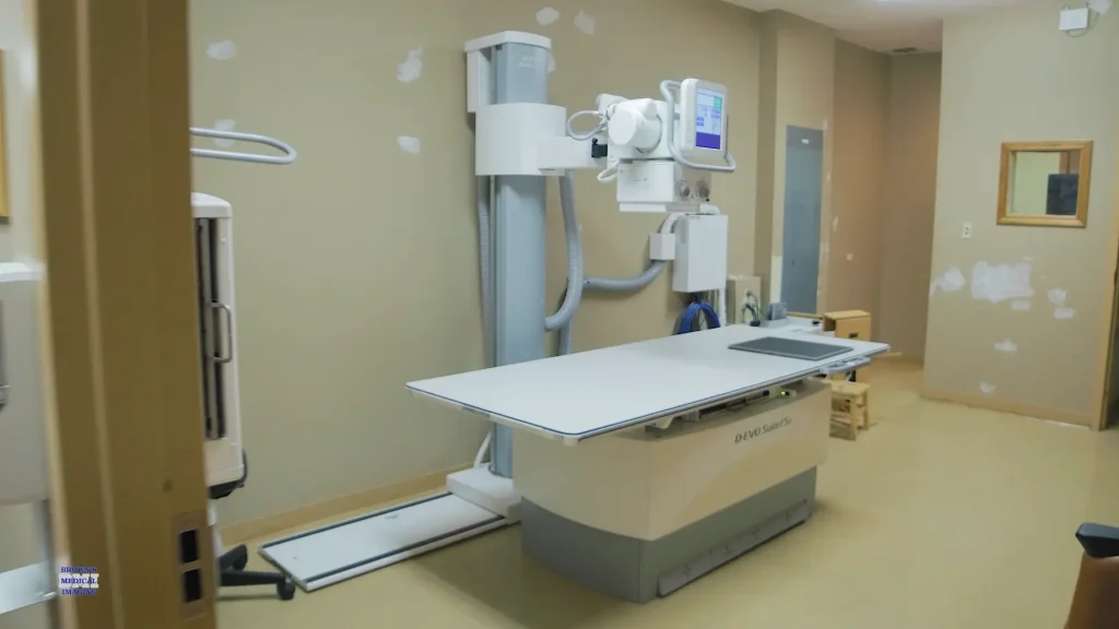
For our smaller clinic, moving to a more advanced imaging system has brought immediate benefits to both patients and staff. Enhanced Patient Comfort & Accessibility The new system features a high–low table, making it easier to assist patients onto the table and position them properly for exams. From helping patients get on and off the […]
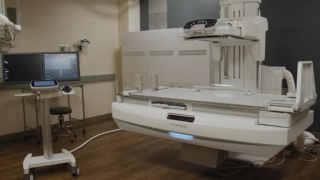
The Fluoro Speed X1 is the latest RF offering from Shimadzu Medical Systems, combining advanced imaging capabilities with thoughtful ergonomic design. Paired with Brown’s Medical Imaging’s service and support, it’s a system built to deliver dependable performance for both patients and technologists. Seamless Integration and Smart Workflow The X1 integrates with multiple DR platforms, allowing […]
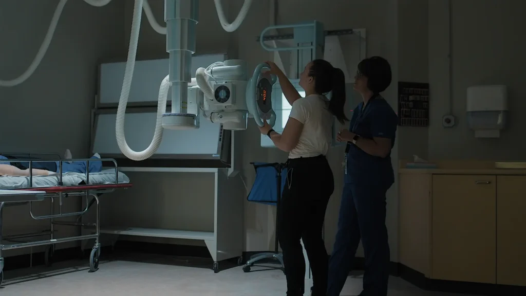
At Clarkson College, hands-on learning is essential for preparing students to excel in their clinical rotations and future careers. That’s why the college has partnered with Brown’s Medical Imaging — giving students access to state-of-the-art diagnostic equipment before they ever step into a hospital or clinic. Practice on the Technology They’ll Use in the Field […]
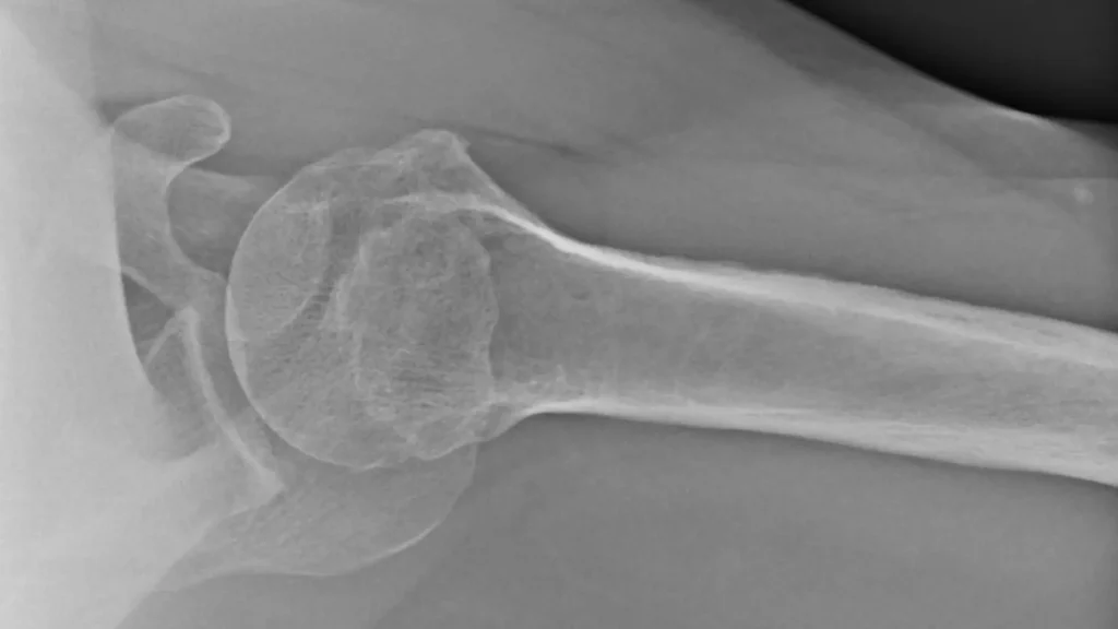
The PA axillary shoulder view is often performed with the patient lying supine on the table. However, on certain digital systems, this projection can be done with the patient sitting or standing, which is easier for both technologist and patient. Advantages of the Upright Method No need for the patient to lie down — ideal […]
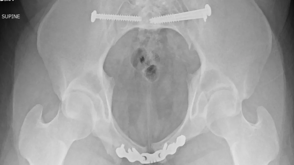
Performing inlet and outlet pelvic views is much simpler on a U-arm X-ray system compared to traditional table-based positioning. With the U-arm, patients can remain standing, eliminating the need for complicated tube angulation while lying down. Inlet View Have the patient stand facing away from the detector. Maintain the built-in caudal angle on the U-arm. […]
